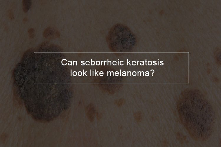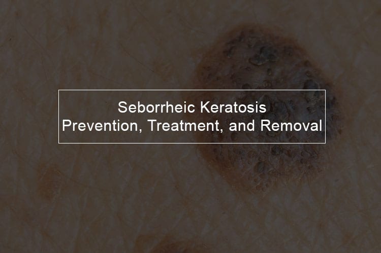
Seborrheic keratosis, also known as seborrheic verruca are benign skin tumors which resemble melanoma. Similar in symptoms to liver spots, seborrheic keratosis develops in the outermost layer of the skin. Seborrheic tumors can vary by location, size, and appearance, but are light tan to dark brown. In this post we will discuss seborrheic keratosis; its causes, symptoms, treatment and how to prevent it. Read through to know more about seborrheic keratosis.
Can seborrheic keratosis appear suddenly?
Seborrheic keratosis causes
Seborrheic Keratosis is limited to discolored skin lesions that occur to be stuck on the epidermis. These patches may take shape suddenly, may vary in size and tend to grow slowly.
The real cause of Seborrheic keratosis is unknown even though researchers believe some forms may be inherited as a dominant trait. Human personalities including the classic genetic diseases are the product of the interaction of two genes for the condition- one received from the father and one from the mother.
In prevailing conditions, a single copy of the disease- either received from either the mother or father- will be expressed dominating the normal gene and resulting in the occurrence of the illness. The threat of transmitting the disorder from affected parent to offspring is 50 percent for every pregnancy nevertheless of the sex of the resulting child.
Seborrheic keratosis Itchy
Usually, Seborrheic keratosis becomes inflamed or irritated, causing pain and itching. An injury to a seborrheic keratosis can cause an infection. Other signs of seborrheic keratosis may include; round or oval shaped, and vary in color from tan, yellowish-brown to black.
Seborrheic keratosis may also be widespread over the trunk, back and or shoulders. Some may be big enough to be called giants while others may be limited to a small area such as the cheeks. Besides the skin lesions may be waxy, scaling or crusted. They tend to become darker and more extensive with age.
What does seborrheic keratosis look like?
Seborrheic keratosis vs melanoma
Even though seborrheic is not cause for concern, its look-alike melanoma- is a possibly deadly type of skin cancer. Thus, it is essential to understand the difference between the two. Here is what you need to know.
How Seborrheic keratosis growths differ from those of melanoma?
- Seborrheic keratosis is round or oval shaped while those of melanoma might have sides which do not match in size or shape.
- Seborrheic keratosis growths can also be light tan while those of melanoma have a fuzzy border or ragged or blurred edges.
- Unlike melanoma growths, Seborrheic growths have a waxy or scaly surface
- Seborrheic keratosis growths may be capped or stick above the surface and frequently appear in groups of two or core.
- Melanoma growths have a variety of colors within the same mole, have a smooth surface and may bleed or ooze.
- Melanoma growths unlike those of seborrheic may change the color, shape or size over time.
Similarities of seborrheic keratosis and melanoma growths?
1. Both seborrheic keratosis and melanoma growths can be brown or black
2. Beware that like melanoma growths; seborrheic keratosis growths can vary in size.
3. Both seborrheic keratosis and melanoma growths can appear anywhere on the body.
Does the same thing cause seborrheic keratosis and melanoma?
As discussed in the previous sector researchers are not sure of what causes seborrheic keratosis. However, it does seem to run in the families so that genetics might be involved. Unlike melanoma, seborrheic keratosis does not seem to relate to sun exposure.
Exposure to ultraviolet light (UV) from natural sunlight or tanning beds is a leading cause of melanoma. The UV rays destroy the DNA in the skin cells, leading them to become cancerous. With proper sun protection, this might be preventable.
Heredity also contributes to likeliness to get melanoma. You are twice likely to develop the disease if a parent or a sibling has previously been diagnosed with melanoma. Besides, only about one in every ten people diagnosed with melanoma also have a family member who has the disease — most of the melanoma diagnoses related to sun exposure.
Can seborrheic keratosis change color?
Seborrheic keratosis is usually brown, but they may also be yellow, white or black. Many lesions may develop, with time; even though at the beginning it might just be one lesion. Patches can be found on many areas of the body, including the: chest, scalp, shoulders, back, abdomen and face. Seborrheic keratosis growths can be found anywhere on the body except on the soles of the feet or the palms.
Can you pick off seborrheic keratosis?
Is it ok to pick off seborrheic keratosis?
Most doctors do not encourage the practice, but you can flick off a seborrheic keratosis. Sometimes you might get it scrapped off by accident. The only risk involved is a little bleeding. If seborrheic keratoses are treated, it is basically for cosmetic reasons. Most of the patients use ice to freeze them off. The process is the same as wart removal but quicker. They can be scrapped or shaved off with a particular cutting instrument. However, seborrheic keratosis removal sometimes creates an area with less or no pigment, so it looks white.
Can seborrheic keratosis go away by itself?

Seborrheic keratoses treatment
Before we look at seborrheic treatment let's look at when you should see a doctor. Even though seborrheic is not dangerous, you should not ignore growths on your skin. It can be challenging to distinguish between harmless and dangerous growths, since as already discussed something that appears as seborrheic keratosis could be melanoma.
Have a doctor check your skin if:
- You are experiencing a new growth
- You have observed a new change in the appearance of an existing growth
- See a doctor if growth is irritated or painful
- If your growth has regular borders see a doctor
- A seborrheic keratosis usually occur in multiple thus if you have one single growth you should see a doctor
- See a doctor if your growth has an unusual color, such as purple or blue.
- If you are concerned about any growth, make an appointment with your physician. It' is better to be too cautious than ignore a potentially serious problem.
Seborrheic keratoses, typically do not go away by themselves, but they can be removed if they become irritating. There is no problem in not treating the growths because they are not cancerous and they do not become cancerous. For this reason, most doctors prefer leaving the condition alone.
Apart from irritation, the other exception to when this condition should be treated is if multiple seborrheic keratoses appear suddenly. If this happens, it might be a sign of a tumor growing inside your body. The doctor will test for any underlying conditions and work with you on any steps.
A dermatologist will frequently be able to diagnose seborrheic keratosis by eye. If there’s any uncertainty, they] will likely remove part or all of the growth for testing in a laboratory. This is known as a skin biopsy.
The biopsy will be assessed under a microscope by a trained doctor. This can assist your doctor to diagnose the growth as either seborrheic keratosis or cancer (such as malignant melanoma).
Among the treatment options include
- Burning the tissue with an electric current. The process a local anesthetic or a numbing agent. After the lesion is gotten rid off, it can take at least two or four weeks for the scab to drop and the underlying tissue to heal.
- Seborrheic keratosis may also be treated through liquid nitrogen. In this procedure, the lesion is exposed to 321-degree temperatures, is also well suited for removing smaller lesions. The exposed tissue will rapidly form a blister that crusts over and falls off after some days.
- Shave an excision is also a reasonable option for seborrheic keratosis growths removal. Using a razor and local anesthetic, the growth is shaved off, and the wound is covered with aluminum chloride or silver nitrate mixture to prevent loss of blood. To reduce the appearance of the scar electrocautery may be used to feather the edges.
- Electrodesiccation and curettage is a procedure commonly used to treat seborrheic keratosis growths if they become cancerous. It requires the sweeping of the cancerous tissue with a device known as a curette, followed by the electrocautery to damage the underlying cells. It is performed under local anesthesia and repeated three times to make sure all cancer cells are removed.
If a treatment is performed for cosmetic reasons, extra care needs to be taken for people with darker skin as the removal of a lesion may leave a visibly lighter scar.
Seborrheic keratosis removal
Doctors may decide to remove Seborrheic keratosis growths that have a suspicious appearance or cause physical or emotional discomfort. Most of the conventional methods used in seborrheic keratosis removal are cryosurgery, electrosurgery, and curettage as discussed above.
After the seborrheic keratosis removal, the skin of the patient may be lighter at the site of extraction. The difference in skin color frequently becomes less noticeable over time. Most of the time a seborrheic keratosis will not return, but it is possible to develop a new one on another part.
Tips for prevention of seborrheic keratosis and melanoma
Both seborrheic keratosis and melanoma have been associated with sun exposure. The most effectual method to reduce the risk for either condition is to stay away from tanning beds and protect your skin from the sun. Here are measures which could help to prevent the occurrence of either disease:
- Apply sunscreen with a higher SPF every day
- Applying sunscreen every two hours and also immediately after sweating heavily or swimming
- Avoid being in direct sunlight between 10 a.m and 4 p.m because the sun rays are most penetrating at this time.
- Watch for changes in any existing moles. If you see anything unusual schedule visit a doctor.
Seborrheic keratosis misdiagnosis
To diagnose seborrheic keratosis, a doctor begins by examining the surface characteristics of your growth with a magnifier.
Even though there are visual differences between seborrheic keratosis and melanoma the two conditions, can be misleading. Melanomas sometimes mimic features of seborrheic keratosis so successfully that misdiagnoses are possible. If there is any doubt, the doctor should take a sample of the mole and take it to the laboratory for testing.
Advanced diagnostic tests, such as reflectance confocal microscopy, do not need a Moleskine sample. This type of test uses a special microscope to perform a noninvasive examination. The mechanism is widely used in Europe and is becoming available in America.
It is essential to differentiate the conditions because misdiagnosis might have untoward implications for the patient, as both diagnostic procedures and treatments may be suboptimal.
Who at risk of developing seborrheic keratosis?
Risk factors of seborrheic keratosis include are not limited to
- Old age- the condition frequently occurs in those who are middle age. The risk of seborrheic occurrence increases with age.
- Family members with seborrheic keratosis; the skin problem frequently runs in families. The risk increases with the number of affected relatives.
- Too much exposure to the sun; there is evidence that skin exposed to the sun is more likely to get a seborrheic keratosis. Nevertheless, growths also appear on skin that is usually covered up when people go out in the sun.


